

General Ultrasound
Ultrasound is a safe and non-invasive scan that uses sound waves to create real-time images of the inside of your body. It can be used to examine organs, soft tissues and blood flow, and is often recommended to investigate pain, swelling, lumps or unusual test results. Ultrasound does not use radiation and is generally a comfortable examination.
Available At
What does an ultrasound examine?
Ultrasound can be used to examine many areas of the body, including the abdomen, pelvis, kidneys, thyroid and soft tissues. Because the images are produced in real time, ultrasound can also show movement and blood flow.
Why might I need an ultrasound?
Your doctor may refer you for an ultrasound to investigate pain, swelling, a lump, abnormal blood test results or symptoms affecting a particular organ or area of the body. Ultrasound may also be used to assess blood flow or guide certain medical procedures.
What can I expect?
Your sonographer will apply water-based gel to your skin and gently move a small handheld device, called a transducer, over the area being examined.
Some pressure may be needed to capture clear images. The scan is generally painless, although you may feel some discomfort if the area is already sore or tender. Please let your sonographer know if you are uncomfortable at any time.
How should I prepare?
Preparation will depend on the type of ultrasound you are having. You may be asked to avoid eating for a certain period, drink water and arrive with a full bladder, or follow other specific instructions.
Please follow the preparation instructions provided when you book. If you are unsure about anything, please contact our team before your appointment.
Are there any risks involved with an ultrasound?
Ultrasound does not use radiation and is not known to have any harmful effects. Please advise the sonographer if you have any allergies to latex, or any skin sensitivities.
Benefits of ultrasound imaging
Ultrasound does not use radiation and provides real-time images of the body. It can give your doctor valuable information to help make a diagnosis and recommend the most appropriate care.
FAQs
-
No. Ultrasound uses sound waves to create images and does not expose you to radiation.
-
Ultrasound is usually painless. Your sonographer may need to apply some pressure to obtain clear images, which can feel uncomfortable if the area is already tender.
-
Food, gas and the amount of fluid in your bladder can affect how clearly certain organs can be seen. Following the preparation instructions helps your sonographer capture the best possible images.
-
Most ultrasound examinations take around 20 to 30 minutes. This may vary depending on the area being examined and the complexity of the scan.
Pregnancy and
Gynaecological Ultrasound
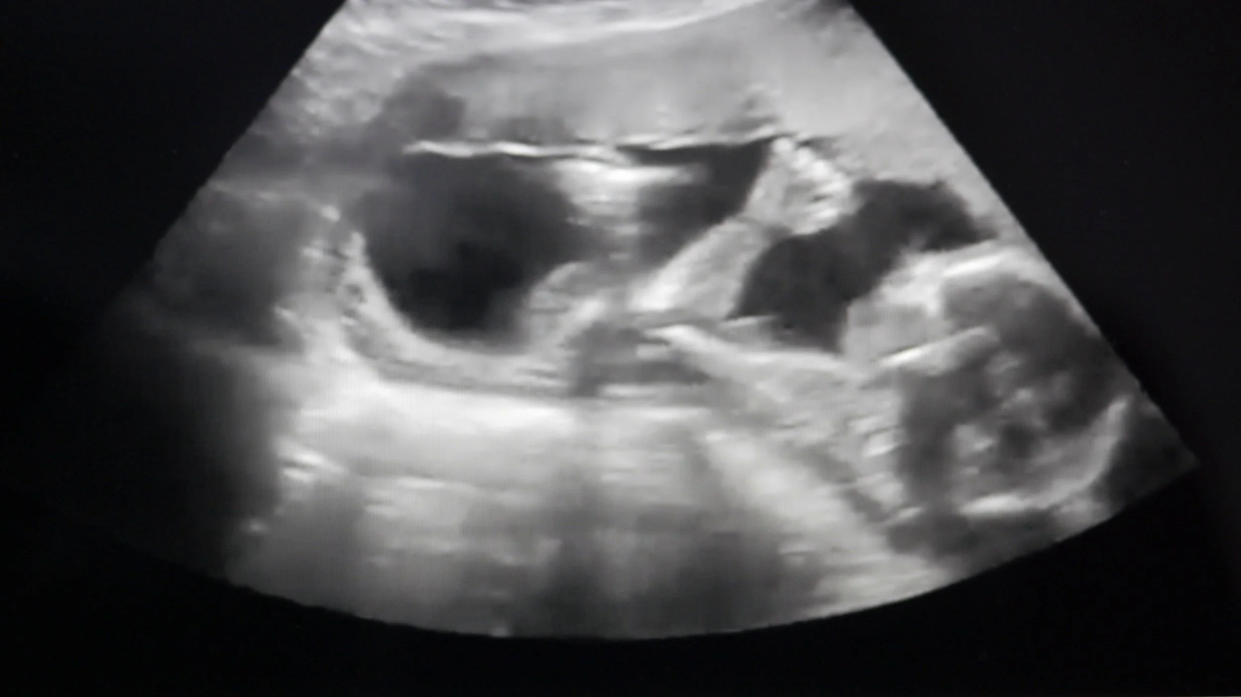
Pregnancy and gynaecological ultrasound provide detailed images of the uterus, ovaries and surrounding pelvic area. During pregnancy, it is also used to check your baby’s growth and development. Your doctor may recommend an ultrasound to investigate pelvic symptoms, fertility concerns or to support your care throughout pregnancy.
Available At
What does the ultrasound examine?
A gynaecological ultrasound looks at the uterus, ovaries and other structures within the pelvis. During pregnancy, ultrasound is used to assess your developing baby and the surrounding structures, including the placenta and amniotic fluid.
Depending on the reason for your scan and the stage of your pregnancy, an ultrasound may be performed over your abdomen, internally, or sometimes using both methods.
Why might I need this ultrasound?
Your doctor may refer you for a gynaecological ultrasound to investigate pelvic pain, unusual bleeding, menstrual concerns, ovarian cysts, fibroids or fertility-related issues.
During pregnancy, an ultrasound may be used to confirm your baby’s due date and position, check growth and development, assess the placenta and amniotic fluid, or look into a specific concern.
What can I expect?
For a pelvic ultrasound, your sonographer will apply water-based gel to your lower abdomen and move a small handheld device across your skin to capture the images.
In some cases, a transvaginal ultrasound may be offered to provide clearer and more detailed images. This involves gently inserting a covered and lubricated ultrasound transducer into the vagina. Your sonographer will explain the examination beforehand, answer any questions and ask for your consent. You can pause or stop the examination at any time.
How should I prepare?
For some pelvic and early pregnancy ultrasounds, you may be asked to drink water and arrive with a full bladder. Preparation depends on the type of scan, so please follow the instructions provided when booking your appointment.
Benefits of pregnancy and gynaecological ultrasound
These ultrasounds provide clear, real-time images that can help your doctor assess gynaecological concerns and support your care throughout pregnancy.
FAQs
-
This will depend on the reason for your scan and the type of images required. Sometimes both methods are helpful. A transvaginal ultrasound will only be performed after it has been explained to you and you have provided your consent.
-
A full bladder is often required for pelvic and early pregnancy ultrasounds. Please follow the preparation instructions provided when booking your appointment, as these may differ depending on your examination.
-
Ultrasound is widely used during pregnancy when clinically required and performed by our highly trained sonographers.
-
You may be able to bring a support person, depending on the clinic and the type of examination. Please check with our team when booking, particularly if you would like to bring children.

Musculoskeletal and Sports Ultrasound
Musculoskeletal ultrasound is used to examine muscles, tendons, ligaments, joints and other soft tissues. It can help identify the cause of pain, swelling or restricted movement and is commonly used to assess sporting injuries. Because the images are produced in real time, the area can often be examined while you move.
Available At
What does it examine?
Musculoskeletal ultrasound provides detailed images of muscles, tendons, ligaments, joints, bursa and other soft tissues close to the skin. Your sonographer can examine the area while it moves and, when helpful, compare it with the same area on the opposite side of your body.
Why might I need this ultrasound?
Your doctor may refer you for a musculoskeletal ultrasound to investigate pain or an injury affecting the shoulder, elbow, wrist, hand, hip, knee, ankle or foot. It may also be used to check for tendon injuries, bursitis, fluid build-up, soft-tissue lumps and other sports-related concerns.
What can I expect?
Your sonographer will apply water-based gel to your skin and move a small handheld device over the area being examined.
You may be asked to change position or perform a few gentle movements so the muscles, tendons or joints can be seen in action. Some pressure may be needed to capture clear images, which can feel uncomfortable if the area is already sore. Please let your sonographer know if you are experiencing pain.
How should I prepare?
There is usually no preparation required. Wear comfortable clothing that allows the area to be reached easily and bring any previous scans or reports if requested.
Benefits of musculoskeletal ultrasound
Musculoskeletal ultrasound provides focused, real-time imaging without using radiation. The results can help your doctor understand the cause of your symptoms, plan your treatment and determine whether an image-guided procedure may be suitable.
FAQs
-
Common areas include the shoulder, elbow, wrist, hand, hip, knee, ankle and foot. Ultrasound can also be used to examine muscles, tendons, bursa and soft-tissue lumps.
-
Yes. One of the main benefits of musculoskeletal ultrasound is that your sonographer can often examine tendons, joints and other structures while you perform gentle movements.
-
Usually, no preparation is needed. We recommend wearing loose, comfortable clothing that allows easy access to the area being examined.
-
An injection can only be performed if you have an appropriate referral and the procedure has been assessed and booked in advance. A diagnostic ultrasound appointment does not automatically include an injection.
Vascular Ultrasound
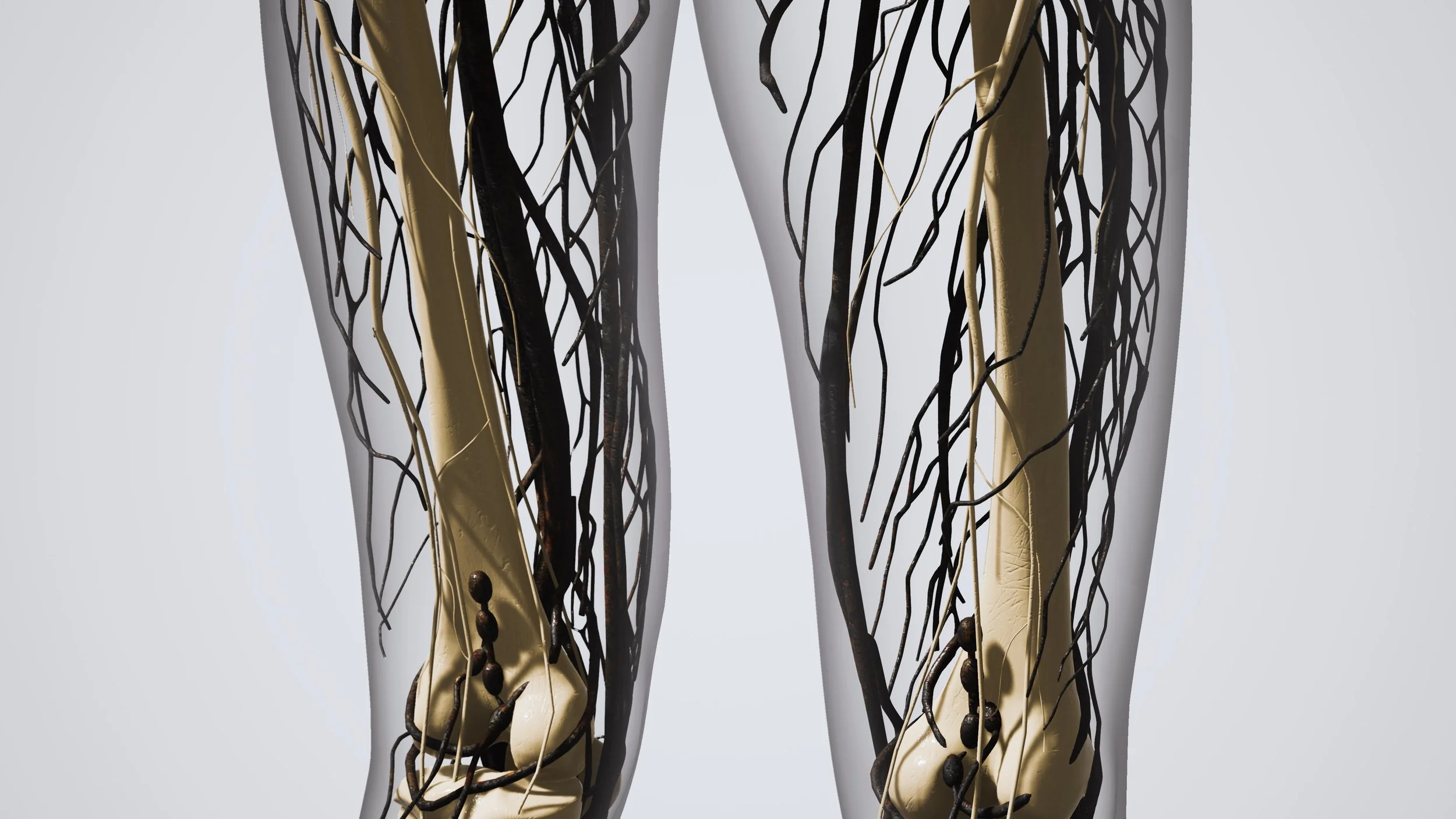
A vascular ultrasound looks at how blood is flowing through the arteries and veins in your body. Your doctor may recommend this scan to investigate pain, swelling, circulation problems, blood clots or narrowing of the blood vessels. It is a non-invasive examination that does not use radiation.
Available At
What does a vascular ultrasound examine?
Vascular ultrasound, also known as Doppler ultrasound, provides images of your blood vessels and shows the direction and speed of your blood flow. It can be used to examine arteries and veins in different areas of the body, including the neck, abdomen, arms and legs.
Why might I need a vascular ultrasound?
Your doctor may refer you for a vascular ultrasound if you have swelling or pain in your arms or legs, varicose veins or symptoms of poor circulation. It is also commonly used to check for deep vein thrombosis (DVT), narrowing of an artery or an aneurysm.
The scan may also be recommended to monitor an existing condition or check your progress after vascular treatment.
What can I expect?
Your sonographer will apply water-based gel to your skin and move a small handheld device over the blood vessels being examined.
Some pressure may be needed, particularly when checking that a vein compresses normally. This may feel uncomfortable if the area is already swollen or tender. You may also hear pulsing sounds from the ultrasound machine as it measures your blood flow.
How should I prepare?
Preparation will depend on the part of your body being examined. Some vascular ultrasounds of the abdomen require fasting, while most scans of the arms and legs need no preparation.
Please follow the instructions provided when booking or contact our team if you are unsure.
Benefits of vascular ultrasound
Vascular ultrasound provides detailed information about your blood flow and circulation without the need for radiation or surgery. The results can help your doctor make a diagnosis and plan or monitor your treatment.
FAQs
-
Doppler ultrasound is a technique used to show the direction and speed of blood flowing through your arteries and veins.
-
Yes. Vascular ultrasound is commonly used to check for suspected blood clots, including deep vein thrombosis.
-
Your sonographer may need to apply firm pressure or gently compress the veins during the scan. This can feel uncomfortable if the area is swollen or sore, but please let your sonographer know if you are experiencing pain.
-
Some vascular ultrasounds of the require fasting, but many scans of the arms and legs do not. Please follow the preparation instructions provided for your specific examination when booking your appointment.

Paediatric Hip Ultrasound
A paediatric hip ultrasound is a safe and non-invasive examination used to assess the development and stability of a baby’s hip joints. It is most commonly performed to check for developmental dysplasia of the hip (DDH), a condition where the hip joint has not developed normally or the top of the thigh bone does not sit securely within the hip socket. Ultrasound provides detailed images of the developing hip without using radiation.
Available At
What a paediatric hip ultrasound can show
The ultrasound allows the sonographer to assess the shape and position of the hip joint and how securely the top of the thigh bone sits within the hip socket. Because babies’ hip joints are still developing and contain a large amount of cartilage, ultrasound is particularly useful for examining the hips during early infancy.
Why paediatric hip ultrasounds are performed
Your doctor or healthcare professional may refer your baby for a hip ultrasound if there is a concern about how the hips are developing. Common reasons include breech presentation, a family history of developmental dysplasia of the hip, an unstable or clicky hip during examination, reduced movement of one hip, uneven skin folds or another clinical concern identified during a routine examination.
What to expect during your baby’s ultrasound
Your baby will usually lie on their back or side while the sonographer gently examines each hip using a small ultrasound transducer and water-based gel. The sonographer may gently move your baby’s legs into different positions so the hip joints can be assessed from several angles. The examination is non-invasive and does not use radiation. Your baby may become unsettled from being positioned or having their legs moved, but the scan itself should not be painful.
Preparing for a paediatric hip ultrasound
There is usually no special preparation required. Dress your baby in clothing that is easy to remove from the waist down and consider bringing a spare nappy, bottle, dummy or other comfort item if your baby uses one. Please bring your baby’s referral and any previous relevant imaging or medical information if requested.
Benefits of paediatric hip ultrasound
Ultrasound provides clear, real-time images of a baby’s developing hip joints without exposing them to radiation. Early identification of developmental hip abnormalities allows your baby’s doctor or specialist to determine whether further assessment, monitoring or treatment is required.
FAQs
-
Your baby may have been referred because of something identified during a routine hip examination or because they have a recognised risk factor for developmental dysplasia of the hip, such as breech presentation or a family history of DDH.
-
DDH is a condition where a baby’s hip joint has not developed normally. The top of the thigh bone may not sit securely within the hip socket, and the severity can vary between babies.
-
The examination is generally painless. Your baby’s legs will need to be gently positioned and moved during the scan, so some babies may become unsettled.
-
Hip ultrasound is mainly used during early infancy while the hip joint is still developing. The appropriate timing depends on why the examination has been requested, so please follow the referral and booking instructions provided by your healthcare professional.
Shear Wave Elastography Ultrasound
Shear Wave Elastography (SWE) is a specialised ultrasound technique used to measure the stiffness of liver tissue. It is sometimes referred to as liver elastography, ultrasound elastography or a liver stiffness scan.
Increased liver stiffness can be associated with fibrosis or scarring of the liver. Shear wave elastography provides a non-invasive way to assess liver stiffness without using radiation.
Available At
What shear wave elastography can show
Shear wave elastography measures how quickly ultrasound-generated shear waves travel through the liver. Stiffer liver tissue generally allows the waves to travel differently, providing a measurement that can help assess the presence and severity of liver fibrosis.
The results are interpreted alongside your other imaging, blood tests and clinical information.
Why shear wave elastography is performed
Your doctor or specialist may recommend liver elastography if there is a concern about chronic liver disease or possible liver fibrosis.
It may be requested for people with conditions or risk factors such as fatty liver disease, abnormal liver function tests, chronic hepatitis or other causes of ongoing liver inflammation or damage. It can also be used to monitor liver stiffness over time.
What to expect during your examination
The examination is similar to a standard abdominal ultrasound. You will usually lie on your back with your right arm raised while the sonographer places ultrasound gel and a transducer over the right side of your abdomen.
You may be asked to briefly hold your breath while several liver stiffness measurements are taken. The examination is non-invasive and should not be painful.
Preparing for shear wave elastography
Fasting is generally required before liver elastography because eating can affect liver stiffness measurements.
Please follow the preparation instructions provided when your appointment is booked, as the required fasting period may vary. Continue your usual medications unless you have been instructed otherwise.
Benefits of shear wave elastography
Shear wave elastography provides a non-invasive measurement of liver stiffness without using ionising radiation.
It can provide useful information about liver fibrosis, help monitor changes over time and, in some circumstances, may reduce the need for more invasive assessment such as liver biopsy.
FAQs
-
Shear wave elastography is a specialised ultrasound technique that measures liver stiffness. Increased stiffness may be associated with fibrosis or scarring of the liver.
-
Not exactly. A standard ultrasound shows the appearance and structure of the liver, while shear wave elastography provides an additional measurement of liver stiffness. The examinations may be performed together depending on your referral.
-
Shear wave elastography can help assess liver stiffness, which may indicate fibrosis. The result is considered together with your medical history, blood tests and other investigations rather than being interpreted on its own.
-
Fatty liver disease can sometimes be associated with inflammation and fibrosis. Your doctor may request liver elastography to assess whether there are signs of increased liver stiffness or scarring.
-
No. It uses ultrasound and does not expose you to ionising radiation.
-
No. The examination is generally painless. You may feel gentle pressure from the ultrasound transducer against the skin.
-
Yes, fasting is generally required because eating can affect liver stiffness measurements. Please follow the specific preparation instructions provided when booking.
-
The elastography measurements themselves are usually relatively quick. Your appointment may take longer if liver elastography is performed as part of a complete abdominal or liver ultrasound.
-
For some patients, elastography can provide enough information about liver stiffness to reduce the need for a biopsy. However, it does not replace biopsy in every situation. Your doctor or specialist will determine which investigations are appropriate for you.